The Chromozone / IAL Biology / Unit 1 / Topic 1
Revision 1B: mammalian transport and cardiovascular disease
Revision for Topic 1B, to work through on your own before the end-of-section test. There is one screen for each spec point: key words, the key facts and a common mistake, then a one-minute "Spot the mistake". At the end is a self-check of 23 marks. Type your self-check answers in the boxes and hand them in at the end; the model answers open after you hand in, so you can mark yourself. It takes about 50 minutes; you can split it into two sittings (1.6 to 1.9, then 1.10 to 1.14). Teachers: set this after core practical 2.
Teach these first
This page builds on these lessons. Teach them before students start here:
- 1.6 Why animals need a heart and mass transport
- 1.7 Blood vessels: structure and function
- 1.8 The mammalian heart and the cardiac cycle
- 1.9 Haemoglobin: carrying oxygen and carbon dioxide
- 1.10 How atherosclerosis develops
- 1.11 to 1.13 Blood clotting, CVD risk factors and antioxidants
- 1.14 Core practical 2: the vitamin C content of food and drink
Key words: 1.6 mass transport
- Mass transport
- The flow of a fluid, such as blood, that carries substances in bulk over long distances. The heart is the pump.
- Surface area to volume ratio (SA:V)
- Surface area divided by volume. The bigger the organism, the smaller its SA:V.
- Diffusion distance
- How far a substance has to diffuse to reach the cells that need it.
1.6 Why many animals have a heart and circulation
Every cell needs oxygen and glucose and must lose carbon dioxide. Diffusion is fast only over very short distances, so a large, active animal needs a pump and a network of vessels.
| Single cell (Amoeba) | Large, active animal (a mammal) | |
|---|---|---|
| SA:V | Large | Small |
| Diffusion distance | Very short: every part is close to the surface | Long: most cells are far from the body surface |
| Demand for oxygen and glucose | Met by diffusion across the cell surface | High (active and warm), so cells use them quickly |
| How substances reach the cells | Diffusion alone | Mass transport in blood pumped by the heart, then a short diffusion from the capillaries |


Common mistake: writing "a large animal has a small surface area". It has a large surface area but a small surface area to volume ratio.
Spot the mistake (2)
Rewrite this sentence so that it is correct, and say why the original is wrong. (2)
"A whale needs a heart because diffusion does not work in large animals."
Check your answer
- Diffusion still happens in large animals, but it is too slow over long distances (1)
- A whale has a small SA:V and long diffusion distances, so it needs mass transport (1)
- ACCEPT a small surface area to volume ratio
Key words: 1.7 blood vessels
- Lumen
- The space inside a blood vessel, through which the blood flows.
- Elastic fibres
- Fibres in a vessel wall that stretch when blood pressure rises and recoil when it falls.
- Valve
- Flaps in a vein that close when blood starts to flow backwards, so blood only flows towards the heart.
1.7 Blood vessels: structure and function
| Vessel and structure | How the structure suits its job |
|---|---|
| Artery thick wall of smooth muscle and elastic fibres; narrow lumen; no valves | Carries blood away from the heart at high pressure. Elastic fibres stretch and recoil, which smooths out the flow. |
| Capillary wall one cell thick (endothelium); very narrow lumen: red blood cells pass one at a time | Exchange with the body cells: a short diffusion distance; huge networks give a large surface area; slow flow gives time for exchange. |
| Vein thin wall with little muscle and elastic tissue; wide lumen; valves | Carries blood back to the heart at low pressure. The wide lumen gives little resistance; valves prevent backflow. |
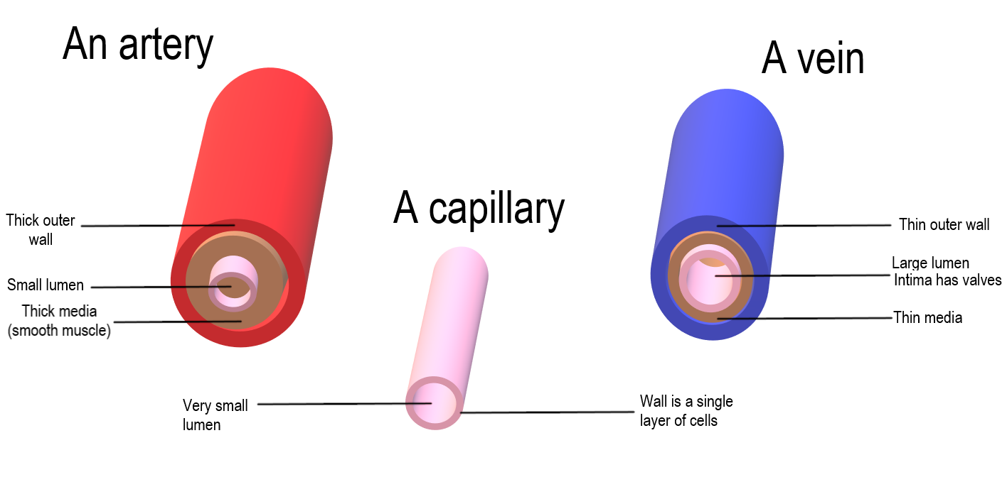

Common mistake: "all arteries carry oxygenated blood". An artery carries blood away from the heart: the pulmonary artery carries deoxygenated blood to the lungs.
Spot the mistake (2)
Rewrite this sentence so that it is correct, and say why the original is wrong. (2)
"The elastic fibres in the walls of arteries contract to pump the blood along."
Check your answer
- Only the heart pumps the blood: elastic fibres cannot contract (1)
- They stretch as pressure rises, then recoil, which smooths the flow (1)
- DO NOT ACCEPT the elastic fibres pump the blood
Key words: 1.8 the heart and the cardiac cycle
- Atrioventricular (AV) valves
- Valves between the atria and the ventricles that stop blood flowing back into the atria.
- Semilunar (SL) valves
- Valves at the bases of the aorta and the pulmonary artery that stop blood flowing back into the ventricles.
- Systole and diastole
- Systole is contraction of heart muscle; diastole is relaxation.
1.8 The heart and the cardiac cycle
Valves open and close only because of differences in blood pressure: blood always flows from higher to lower pressure.
| Stage | Valves and blood flow |
|---|---|
| Atrial systole the atria contract | AV valves open; SL valves closed. Blood is pushed from the atria into the ventricles. |
| Ventricular systole the ventricles contract | AV valves close (ventricle pressure is above atrium pressure); SL valves open (ventricle pressure is above artery pressure). Blood is forced into the aorta and the pulmonary artery. |
| Cardiac diastole all chambers relax | AV valves open; SL valves closed (artery pressure is above ventricle pressure). Blood flows from the veins into the atria and on into the ventricles. |
Major blood vessels: the vena cava brings blood from the body to the right atrium; the pulmonary artery takes it from the right ventricle to the lungs; the pulmonary veins bring it back to the left atrium; the aorta carries it from the left ventricle to the body. The left ventricle has the thickest wall, because it pumps blood all round the body.
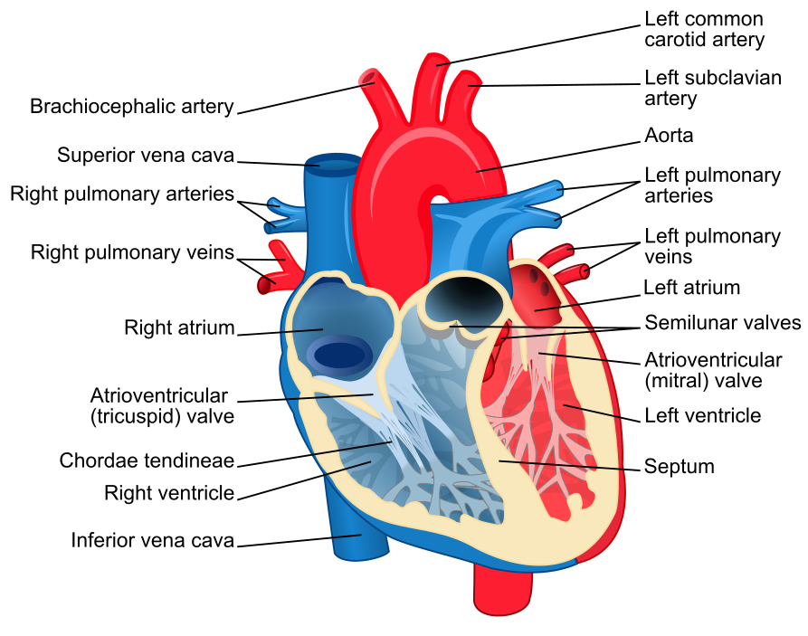

Common mistake: labelling a diagram the wrong way round. A heart is drawn from the front, so its left side is on the right of the picture.
Spot the mistake (2)
Rewrite this sentence so that it is correct, and say why the original is wrong. (2)
"The chordae tendineae pull the AV valves open and shut."
Check your answer
- The AV valves open and close because of pressure differences (1)
- The chordae tendineae stop the valves turning inside out during ventricular systole (1)
- ACCEPT tendons / cords stop the valves being pushed back into the atria
Key words: 1.9 haemoglobin
- Cooperative binding
- When one oxygen molecule binds, haemoglobin changes shape, so the next oxygen molecules bind more easily.
- Bohr effect
- A high concentration of carbon dioxide lowers the affinity of haemoglobin for oxygen, so the curve shifts right and more oxygen is released.
- Fetal haemoglobin
- The haemoglobin of a fetus: it has a higher affinity for oxygen than adult haemoglobin, so its curve lies to the left.
1.9 Haemoglobin and the oxygen dissociation curve
Haemoglobin has four polypeptide chains, each with a haem group holding an iron ion (Fe2+). One molecule carries up to four oxygen molecules.
| Place | pO2 | What haemoglobin does |
|---|---|---|
| Lungs | High (about 13 kPa) | Loads oxygen: about 97% saturated |
| Respiring tissue | Low (2 to 5 kPa) | Unloads oxygen; more where carbon dioxide is high (Bohr effect) |
| Placenta | Low | Fetal haemoglobin takes oxygen from the mother's haemoglobin |
Carbon dioxide travels mostly as hydrogencarbonate ions in the plasma (about 85%), partly bound to haemoglobin (about 10%) and a little dissolved in the plasma.
Common mistake: saying that a curve shifted to the right shows a higher affinity. Right means lower affinity, so more oxygen is released.
Spot the mistake (2)
Rewrite this sentence so that it is correct, and say why the original is wrong. (2)
"In the lungs, where the partial pressure of oxygen is low, haemoglobin loads oxygen until it is about 97% saturated."
Check your answer
- The pO2 in the lungs is high, not low (1)
- At high pO2 haemoglobin loads oxygen readily (1)
- IGNORE haemoglobin is 97% saturated (given in the sentence)
Key words: 1.10 atherosclerosis
- Endothelial dysfunction
- Damage to the endothelium, the inner lining of an artery. It is the first step in atherosclerosis.
- Inflammatory response
- The body's reaction to damage: white blood cells move out of the blood into the damaged tissue.
- Atheroma
- A fatty plaque of cholesterol, white blood cells, fibres and calcium salts in an artery wall, under the endothelium.
1.10 How atherosclerosis develops
Atherosclerosis happens in arteries, where the blood pressure is high. Learn the four steps in order.
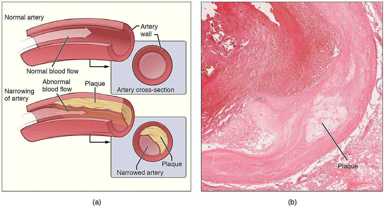

Common mistake: describing an atheroma as fat stuck to the inside of the lumen, like grease in a pipe. It builds up inside the artery wall, under the endothelium.
Spot the mistake (2)
Rewrite this sentence so that it is correct, and say why the original is wrong. (2)
"Atherosclerosis starts when cholesterol sticks to the surface of a healthy, undamaged artery lining."
Check your answer
- It starts with damage to the endothelium (endothelial dysfunction) (1)
- The plaque forms under the endothelium, after an inflammatory response (1)
- IGNORE walls of the blood vessels (it must say endothelium / lining)
Key words: 1.11 blood clotting
- Thromboplastin
- An enzyme released by damaged tissue and activated platelets. It starts the clotting cascade.
- Thrombin
- The active enzyme that converts fibrinogen into fibrin. Its inactive form is prothrombin.
- Fibrin
- An insoluble, fibrous protein. Its strands form a mesh that traps blood cells to make a clot.
1.11 Blood clotting and its role in CVD
Role in CVD: if an atheroma ruptures, the clotting cascade starts and a clot (thrombus) forms inside the artery. In a coronary artery it can cut off the blood supply to part of the heart muscle (a heart attack, or myocardial infarction). In an artery to the brain it causes a stroke.
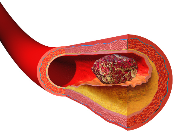

Common mistake: mixing up thromboplastin and thrombin. Thromboplastin turns prothrombin into thrombin; thrombin turns fibrinogen into fibrin.
Spot the mistake (2)
Rewrite this sentence so that it is correct, and say why the original is wrong. (2)
"Prothrombin is an enzyme released by damaged tissue, and it starts the clotting cascade."
Check your answer
- Thromboplastin is the enzyme released by damaged tissue and platelets (1)
- Prothrombin is an inactive, soluble plasma protein that becomes thrombin (1)
- DO NOT ACCEPT prothrombin released by damaged tissue
Key words: 1.12 risk factors
- Risk factor
- Anything that increases the chance of developing a disease. It does not mean the disease will definitely develop.
- Cardiovascular disease (CVD)
- Disease of the heart and blood vessels, such as coronary heart disease and stroke.
1.12 Risk factors for CVD
| Factor | How it raises the risk |
|---|---|
| Genetics | Some people inherit a tendency to high blood pressure or high blood cholesterol. Cannot be changed. |
| Diet | Saturated fat raises blood cholesterol; salt raises blood pressure. Can be changed. |
| Age | Arteries become stiffer and damage builds up over the years. Cannot be changed. |
| Gender | Men have a higher risk than women of the same age until the menopause; oestrogen seems to protect women. Cannot be changed. |
| High blood pressure | Damages the endothelium, the first step of atherosclerosis. Can be lowered by lifestyle or drugs. |
| Smoking | Chemicals in smoke damage the endothelium; nicotine raises blood pressure. Can be changed. |
| Inactivity | Raises blood pressure and makes obesity more likely. Can be changed. |
Each risk factor raises the chance of CVD, and several together raise it more.
Common mistake: writing that a risk factor causes CVD. It only makes CVD more likely.
Spot the mistake (2)
Rewrite this sentence so that it is correct, and say why the original is wrong. (2)
"Women and men of the same age always have the same risk of CVD."
Check your answer
- Women have a lower risk than men of the same age until the menopause (1)
- The hormone oestrogen seems to protect women (1)
- ACCEPT after the menopause the risk becomes similar
Key words: 1.13 antioxidants
- Free radical
- An atom or molecule with an unpaired electron, which makes it highly reactive.
- Antioxidant
- A molecule that makes free radicals stable by donating electrons to them. Vitamin C is one.
- Oxidative stress
- More free radicals in the body than its antioxidants can deal with.
1.13 Dietary antioxidants and the risk of CVD
The evidence: people who eat a lot of fruit and vegetables have less CVD. This is a correlation: they may also exercise more or smoke less. A controlled trial is needed to test whether antioxidants themselves lower the risk.
Common mistake: writing that antioxidants "kill" free radicals or remove cholesterol. They donate electrons, so the free radicals become stable.
Spot the mistake (2)
Rewrite this sentence so that it is correct, and say why the original is wrong. (2)
"Free radicals are stable molecules that protect the lining of arteries."
Check your answer
- Free radicals are highly reactive: they have an unpaired electron (1)
- They damage the endothelium, which can start atherosclerosis (1)
- DO NOT ACCEPT free radicals are stable / protect the artery
Key words: 1.14 core practical 2
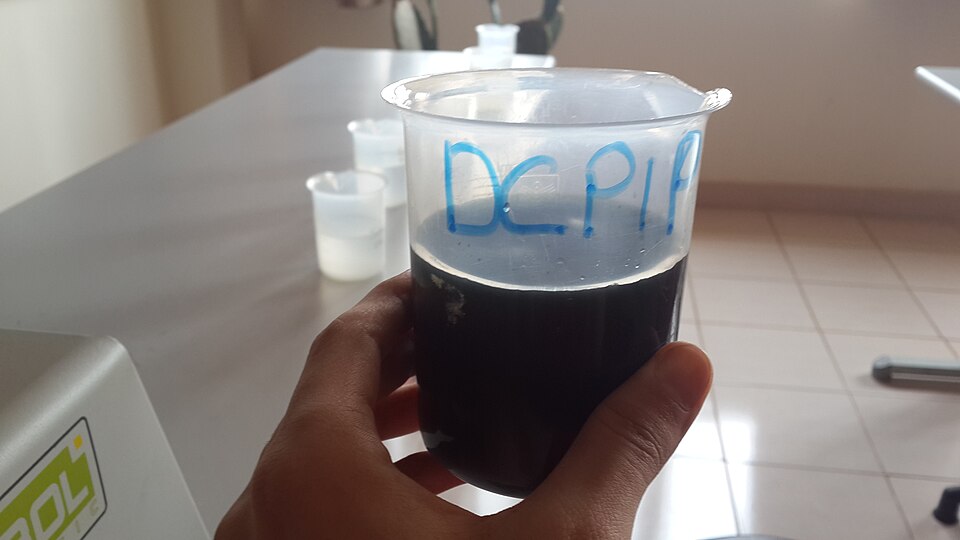
- DCPIP
- A blue dye that becomes colourless when vitamin C reduces it.
- End point
- The moment the last of the blue colour of the DCPIP disappears.
- Standard solution
- A solution whose concentration is known exactly, for example 1.0 mg cm−3 vitamin C.
1.14 Core practical 2: the vitamin C content of food and drink
| Step | What you do |
|---|---|
| 1 | Put the same volume of DCPIP (for example 1.0 cm3) in a test tube. |
| 2 | Add the vitamin C standard drop by drop, shaking, until the blue colour just disappears. Record the volume. |
| 3 | Repeat three times and calculate a mean, leaving out any anomalous result. |
| 4 | Repeat with each juice, using fresh DCPIP each time. |
| 5 | Calculate the vitamin C concentration of each juice (formula below). |
Variables: the independent variable is the type of juice; the dependent variable is the volume needed to decolourise the DCPIP. Keep the volume and concentration of DCPIP the same, and test the juices soon after opening them.

Common mistake: thinking a bigger volume of juice means more vitamin C. The more juice needed, the less vitamin C it contains.
Spot the mistake (2)
Rewrite this sentence so that it is correct, and say why the original is wrong. (2)
"Juice A needed 2.0 cm³ to decolourise the DCPIP and juice B needed 1.0 cm³, so juice A contains more vitamin C."
Check your answer
- Juice B contains more vitamin C (twice as much per cm³) (1)
- A smaller volume of juice B held enough vitamin C to decolourise the DCPIP (1)
- ACCEPT juice A needed more volume, so it is more dilute
Self-check, part A: 1.6 to 1.9 (11 marks)
Answer from memory first, then look back at the screens if you need to. Allow about a minute per mark. The spec point is in brackets after each question number.
1. An earthworm is about 5 mm thick. Oxygen diffuses into its body through its thin, moist skin, yet it also has a heart and blood vessels. Explain why the earthworm needs a circulation even though oxygen enters through its skin. (1.6) (3)
2. Vessel X has a wall 1.0 mm thick, a lumen 4 mm across and no valves. Vessel Y has a wall 0.5 mm thick, a lumen 8 mm across and valves. Identify vessel X and vessel Y, giving one reason for each. (1.7) (2)
3(a) Explain why the wall of the left atrium is much thinner than the wall of the left ventricle. (1.8) (2)
3(b) In one patient, the AV valve on the right side of the heart does not close properly. Suggest what happens to some of the blood during ventricular systole. (1.8) (1)
4. Use the graph in section 1.9. At a pO2 of 4 kPa, read the percentage saturation of adult haemoglobin at low and at high carbon dioxide, and calculate the difference. Explain why this difference helps a muscle during exercise. (1.9) (3)
Model answers for part A
1. The earthworm (3)
- Its body is too thick for diffusion alone to reach the inner cells fast enough (1)
- Blood carries oxygen from the skin to all the cells by mass transport (1)
- Blood carries oxygen away, so a steep gradient at the skin is kept (1)
- ACCEPT long diffusion distance / small SA:V for the first point
2. Vessels X and Y (2)
- X is an artery: thick wall, narrow lumen or no valves (1)
- Y is a vein: thinner wall, wide lumen or valves (1)
- NB both identifications need a reason from the vessel structure
3(a) The left atrium (2)
- The atrium only pumps blood a short way, into the ventricle (1)
- The ventricle pumps blood all round the body at high pressure (1)
- ACCEPT the ventricle wall is thicker / has more muscle
3(b) A faulty AV valve (1)
- Blood flows back into the right atrium (backflow) (1)
- DO NOT ACCEPT blood flows into the left atrium
4. Reading the curves (3)
- Readings: about 57% at low CO2 and 38% at high CO2 (1)
- Difference about 19 percentage points (accept 16 to 22) (1)
- Exercising muscle makes more CO2, so more oxygen is released for respiration (1)
- ACCEPT readings within 3% of 57% and 38%
- ALLOW ecf from the readings for the difference
Self-check, part B: 1.10 to 1.14 (12 marks)
5. Put these events in the order in which they happen in atherosclerosis. Write the letters. A white blood cells move into the artery wall; B high blood pressure damages the endothelium; C the lumen narrows and blood pressure rises; D cholesterol, calcium salts and fibres build up under the endothelium; E more areas of endothelium are damaged. (1.10) (2)
6. A patient's blood contains much less fibrinogen than normal. Explain why a small cut bleeds for longer than usual. (1.11) (2)
7. Person P is a man aged 62 who smokes and whose father had a heart attack at 55. Person Q is a woman aged 35 who does not smoke and has normal blood pressure. State which person has the higher risk of CVD, and give two reasons from this information. (1.12) (2)
8(a) In a survey, people who ate the most fruit and vegetables had fewer heart attacks than people who ate the least. Suggest one other difference between the two groups that could explain this result. (1.13) (1)
8(b) A free radical takes an electron from a molecule in the membrane of an endothelial cell. Suggest how this could lead to an atheroma. (1.13, 1.10) (2)
9(a) 1.0 cm3 of DCPIP was decolourised by a mean of 0.50 cm3 of a 1.0 mg cm−3 vitamin C standard, and by a mean of 2.5 cm3 of orange juice. Calculate the vitamin C content of the orange juice in mg per 100 cm3. (1.14) (2)
9(b) Give one reason why the same test on a dark red berry juice may be less accurate. (1.14) (1)
Model answers for part B
5. The order (2)
- B, A, D, C, E: all five in the right order (1)
- NB one mark if only one pair is swapped
6. Less fibrinogen (2)
- Thrombin has less fibrinogen to convert into fibrin (1)
- Less insoluble fibrin mesh forms to trap cells, so the clot forms slowly (1)
- DO NOT ACCEPT fibrin converted into fibrinogen
7. Two people (2)
- Person P, with any one reason: older, male, smokes, or family history (1)
- A second reason from that list (1)
- NB both marks need person P
- NB person Q is younger, a woman before the menopause, and does not smoke
8(a) Another difference (1)
- For example: they exercise more, smoke less or eat less saturated fat (1)
- ACCEPT any other lifestyle or genetic difference linked to lower CVD risk
8(b) Free radicals and atheroma (2)
- The endothelium is damaged (endothelial dysfunction) (1)
- An inflammatory response follows: white blood cells enter the wall and plaque builds up (1)
- IGNORE walls of the blood vessels (it must say endothelium / lining)
9(a) The calculation (2)
- 0.50 mg of vitamin C in 2.5 cm3, so 0.20 mg cm−3 (1)
- = 20 mg per 100 cm3 (1)
- NB a correct answer of 20 mg per 100 cm3 with no working scores both marks
- ALLOW ecf from the 0.20 mg cm−3 for the final answer
9(b) The red juice (1)
- Its own colour makes the end point hard to see (1)
- ACCEPT the colour change is masked / cannot see the DCPIP decolourise
RAG self-assessment
Mark your self-check, then decide what to revise before the end-of-section test.
- Red: I scored less than half. I will go back to the lessons for the spec points I got wrong.
- Amber: I know most of the facts, but I lose marks on graphs, calculations or the order of events.
- Green: I scored 18 or more and can explain every answer in my own words.
Next: the end-of-section test
When you are confident with every spec point, your teacher will set the Topic 1B end-of-section test.